Back Muscle Diagram Labeled : Back Of The Head Muscle Structure And Nerve System Diagram Stock Vector Illustration Of Labeled Muscle 171279797 / As these muscles contract and relax, they move skeletal bones to create movement of the body.
byAdmin-
0
Back Muscle Diagram Labeled : Back Of The Head Muscle Structure And Nerve System Diagram Stock Vector Illustration Of Labeled Muscle 171279797 / As these muscles contract and relax, they move skeletal bones to create movement of the body.. In this image, you will find an occipital bone, sternocleidomastoid, trapezius, deltoid in muscles of the lower back diagram. If you're looking for a speedy way to learn muscle anatomy, look no further than our anatomy crash courses. See back muscles and low back pain. 1) above the cervical area (longissimus capitis), 2) in the cervical area (longissimus cervicis), and 3) in the upper back or thoracic area (longissimus thoracis). Muscles of the head and neck anatomy pictures and inform.
Muscles of the head and neck anatomy pictures and inform. This article looks at the anatomy of the back, including bones, muscles, and nerves. These layers of back muscles help to mobilize and stabilize your trunk during your day to day activities. Name the tough connective tissue cord that serves to attach a muscle to a bone. The vertebral column of the lower back includes the five lumbar vertebrae, the sacrum, and the coccyx.
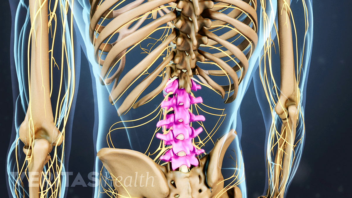
Understanding Lower Back Anatomy from embed.widencdn.net Link to client back care guide The vertebral column of the lower back includes the five lumbar vertebrae, the sacrum, and the coccyx. See back muscles and low back pain. Individual muscle fibers, (b) surrounds groups of skeletal muscle fibers (fascicles), and (c) covers the muscle as a whole. Extrinsic and intrinsic.the back functions are many, such as to house and protect the spinal cord, hold the body and head upright, and adjust the movements of the upper and lower limbs. We hope this picture anatomy of back muscles diagram can help you study and research. Major muscles back muscles shoulder muscles body anatomy human anatomy muscle chart anatomy human muscle anatomy supraspinatus muscle back workout routine. Muscles of lower back diagram.
To learn more about the anatomy of the spine, watch this video.
There are three sets of longissimus muscles: In this image, you will find 1st cervical vertebrae, atlus, cervical plexus, 7th cervical vertebrae, 1st thoracic vertebrae, brachial plexus, spinal dura mater, filaments of spinal nerve roots, 12th thoracic vertebra, 1st lumber vertebra, iliohypogastric nerve, ilioinguinal nerve, lumbar. If you're looking for a speedy way to learn muscle anatomy, look no further than our anatomy crash courses. Claim your free copy of the client back care guide today. Learn vocabulary, terms and more with flashcards, games and other study tools. Muscles of lower back diagram. The bones of the pelvis and lower back work together to support the body's weight, anchor the abdominal and hip muscles, and protect the delicate vital organs of the vertebral and abdominopelvic cavities. Still, many individuals pay far too little attention to them. It also covers some common conditions and injuries that can affect the back. The back is the body region between the neck and the gluteal regions. The muscles of the back can be arranged into 3 categories based on their location: For more anatomy content please follow us and visit. See back muscles and low back pain.
The quadratus lumborum muscles (orange, in the image above) are found in the lower back (also called the lumbar area). As these muscles contract and relax, they move skeletal bones to create movement of the body. Muscles of the head and neck anatomy pictures and inform. Our latest youtube film is ready to run. Link to client back care guide
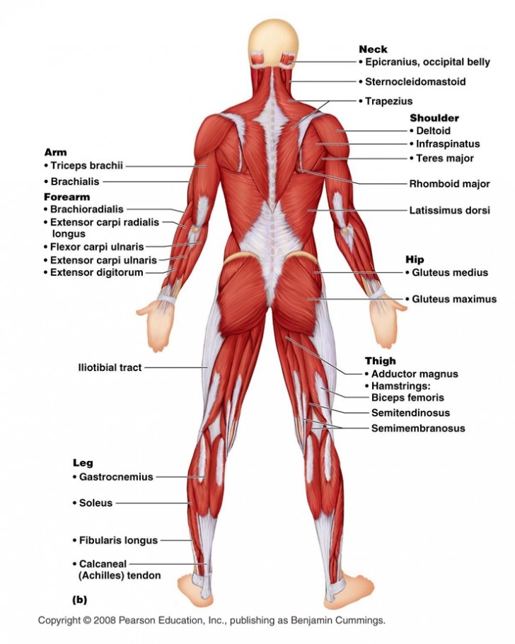
Ucsf Anatomy Back Muscles Ankiweb from dl4.ankiweb.net We are pleased to provide you with the picture named anatomy of back muscles diagram. Muscle diagrams are a great way to get an overview of all of the muscles within a body region. The back anatomy includes some of the most massive and functionally important muscles in the human body. Most of the time, back muscle pain is diagnosed then treated with little more than a prescription of rest, painkillers and muscle relaxants. Individual muscle fibers, (b) surrounds groups of skeletal muscle fibers (fascicles), and (c) covers the muscle as a whole. Name three types of fiber arrangements seen in skeletal muscle. We are pleased to provide you with the picture named labelled diagram of the muscles in the human body.we hope this picture labelled diagram of the muscles in the human body can help you study and research. As you can see, there are also have a spine of scapula deltoid, triceps brachii, latissimus dorsi.
Your back consists of three distinct layers of muscles, namely the superficial layer, the intermediate layer, and the deep layer.
There are three sets of longissimus muscles: The muscles of the lower back help stabilize, rotate, flex, and extend the spinal column, which is a bony tower of 24 vertebrae that gives the body structure and houses the spinal cord.the spinal. Back muscles chart, back muscles diagram and ligaments, back muscles diagram lats, back muscles diagram massage, upper back muscles chart, human muscles, back muscles. Superficial back muscles, intermediate back muscles and intrinsic back muscles.the intrinsic muscles are named as such because their embryological development begins in the back, oppose to the superficial and intermediate back muscles which develop elsewhere and are therefore classed as extrinsic muscles. By the way, have you heard about the myth of. Female reproductive system 2021 | 4 minutes of easy learning mystery female body. Poorly developed back muscles lead to everything. Muscles of the back diagram. Extrinsic and intrinsic.the back functions are many, such as to house and protect the spinal cord, hold the body and head upright, and adjust the movements of the upper and lower limbs. They also attach your shoulders and pelvis to the trunk, creating a bridge between. This article looks at the anatomy of the back, including bones, muscles, and nerves. Muscles of the head and neck anatomy pictures and inform. Claim your free copy of the client back care guide today.
We hope this picture anatomy of back muscles diagram can help you study and research. Related posts of diagram of female back muscles muscle anatomy diagram. These layers of back muscles help to mobilize and stabilize your trunk during your day to day activities. The majority of muscles in the leg are considered long muscles, in that they stretch great distances. The back muscles enable you to stand up straight;
Anatomy Posterior Muscular System Diagram Biological Science Picture Directory Pulpbits Net from pulpbits.net Your clients will thank you for it! Muscle diagram labeled front and back, muscle system labelling (front and back), muscular system labeled front and back, human muscles, muscle diagram labeled front and back, muscle system labelling (front and back), muscular system labeled front and back. 12 photos of the muscles of the lower back and buttocks diagram. Superficial, intermediate, deep and deepest layers.these muscles lie on each side of the vertebral column, deep to the thoracolumbar fascia they span the entire length of the vertebral column, extending from the cranium to the pelvis It also covers some common conditions and injuries that can affect the back. 12 photos of the muscles labeled front and back. Back muscles chart, back muscles diagram and ligaments, back muscles diagram lats, back muscles diagram massage, upper back muscles chart, human muscles, back muscles. Extrinsic and intrinsic.the back functions are many, such as to house and protect the spinal cord, hold the body and head upright, and adjust the movements of the upper and lower limbs.
This article looks at the anatomy of the back, including bones, muscles, and nerves.
Your clients will thank you for it! Support and protect your spine; These layers of back muscles help to mobilize and stabilize your trunk during your day to day activities. Another common cause of lower back and hip pain is disc injury. They also attach your shoulders and pelvis to the trunk, creating a bridge between. The deep back muscles, also called intrinsic or true back muscles, consist of four layers of muscles: Individual muscle fibers, (b) surrounds groups of skeletal muscle fibers (fascicles), and (c) covers the muscle as a whole. The back muscles enable you to stand up straight; In this image, you will find an occipital bone, sternocleidomastoid, trapezius, deltoid in muscles of the lower back diagram. This picture also contains humerus, olecranon process of ulna, deep to tendon and so on. The back is the body region between the neck and the gluteal regions. The muscles, bones, ligaments, and tendons in the back can all be injured and cause back pain. 1) above the cervical area (longissimus capitis), 2) in the cervical area (longissimus cervicis), and 3) in the upper back or thoracic area (longissimus thoracis).